
Peptide chemistry
NAD+ identity: structure, molecular weight and form
NuuVive Journal · · 7 min read

Short answer
Nicotinamide adenine dinucleotide (NAD+) is a dinucleotide coenzyme, not a peptide. The free acid form has a molecular formula of C21H27N7O14P2 and an average molecular weight of 663.43 g/mol. It consists of a nicotinamide riboside monophosphate linked to an adenosine monophosphate via a pyrophosphate bridge. Laboratories typically encounter it as a white, highly hygroscopic lyophilised powder requiring strict cold-chain storage and moisture control during analytical weighing.
Key takeaways
- NAD+ is a dinucleotide coenzyme containing zero amino acids, despite frequently appearing in synthetic peptide catalogues.
- The standard free acid form carries a molecular mass of 663.43 g/mol, which shifts if the material is supplied as a sodium or lithium salt.
- The solid is highly hygroscopic, requiring careful moisture quantification to prevent systematic weighing errors during laboratory preparation.
- Liquid analytical samples degrade rapidly in room-temperature autosamplers and require cooling to yield accurate chromatographic data.
- Mass spectrometry often produces a false sodium adduct peak that analysts mistake for a sodium salt powder.
A peptide in name only
Browse a supplier catalogue for long enough and you will eventually stumble across the phrase "NAD+ peptide". It sits neatly next to the amino acid sequences, sharing the same shelf space and the same glass vials. The label suggests a familiar chain of amide bonds, but the chemistry tells an entirely different story. Nicotinamide adenine dinucleotide contains zero amino acids. The "nicotinamide" part of the name refers to the amide derivative of nicotinic acid, a pyridine ring, rather than any structural link to a peptide chain.
NAD+ is actually a dinucleotide coenzyme. The molecule is built from two distinct nucleotide units, a nicotinamide riboside monophosphate and an adenosine monophosphate. These two halves are stitched together tail-to-tail by a pyrophosphate bridge. It is a complex, fragile architecture designed around a specific reactive site. The structural core responsible for its chemical reactivity is the positively charged pyridinium nitrogen on the nicotinamide ring. This arrangement allows the C4 position to accept a hydride during redox reactions.
Grouping it with synthetic proteins is just a quirk of laboratory supply logistics. Sellers categorise it that way because it ships in the same lyophilised format and requires the same cold-chain handling as actual amino acid chains. But knowing what sits in the vial changes how you prepare a sample. Handle a dinucleotide like a standard synthetic sequence, and the analytical results will quickly spiral out of control.
The molecular weight and formula of the free acid
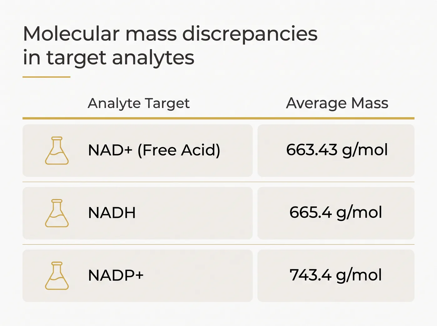
Before you can calibrate an instrument, you need the exact dimensions of the target. The base form of NAD+ is a free acid, acting as an inner salt. Its molecular formula is C21H27N7O14P2, yielding an average molecular weight of 663.43 g/mol and a monoisotopic mass of 663.109 Da. The chemical registry identifies this specific free acid under CAS number 53-84-9. When you open a laboratory report, this is the baseline mass you expect the testing facility to confirm.
Identity testing must also cleanly separate the target from closely related metabolites. Add two hydrogen atoms to the nicotinamide ring, and the mass shifts to 665.4 g/mol. That heavier molecule is NADH. Throw an extra phosphate group onto the adenosine ribose ring, and the scale tips to 743.4 g/mol for NADP+. A careless analytical run might blur these peaks together, but a tight method resolves them.
| Compound | Formula | Average Mass (g/mol) | Structural Difference |
|---|---|---|---|
| NAD+ (Free Acid) | C21H27N7O14P2 | 663.43 | Base dinucleotide |
| NADH | C21H29N7O14P2 | 665.4 | Addition of two hydrogen atoms |
| NADP+ | C21H28N7O17P3 | 743.4 | Addition of a phosphate group |
Salt forms and the trouble with the scale
The mass of the molecule on paper rarely matches the mass of the powder on the balance. Commercially, manufacturers supply NAD+ either as the standard free acid or as an alkali metal salt. A sodium salt version shifts the formula weight up to 685.4 g/mol. That extra 22 g/mol might seem trivial, but it quietly ruins precision work. If a technician weighs the powder assuming it is the free acid, but the vial actually holds the sodium salt, the resulting liquid contains a lower concentration of the active molecule.
Catching this mismatch is harder than it looks. Standard high-performance liquid chromatography (HPLC) relies on ultraviolet light to quantify purity. The problem is that the sodium counter-ion is invisible to UV detection at the standard wavelengths, and it dissociates the moment the powder hits the solvent. The chromatogram will show a single, beautiful peak for NAD+, leaving you entirely blind to the salt form. Verifying the exact composition requires referencing the manufacturer certificate or ordering a separate elemental analysis to check for sodium or lithium.
Catching water: the hidden mass in a lyophilised vial
Salt is not the only invisible variable in the vial. NAD+ is highly hygroscopic, meaning the powder pulls moisture directly out of the surrounding air. The manufacturing process involves a strict freeze-drying step designed to remove water, pushing the material through freezing, primary drying and secondary drying phases. Residual moisture directly affects the physical stability of the dried solid.
If a vial sits open on a laboratory bench on a humid day, the powder acts like a sponge. That trapped water inflates the apparent mass of the sample. Weighing out powder without adjusting for the water content causes a systematic under-calculation of the actual compound present. Laboratories bypass this trap by running a Karl Fischer titration, a technique that quantifies the exact percentage of water locked inside the solid. Good analytical data always accounts for both the salt form and the moisture content before a single calculation takes place.
Laboratory analysis: pushing NAD+ through the column
Getting a clear picture of what is inside the vial requires pushing a liquid sample through an analytical column under high pressure. NAD+ is exceptionally polar. In reversed-phase HPLC, the molecule struggles to bind to standard carbon-coated silica beads. Analysts force it to behave by using carefully optimised mobile phases, often pairing a C18 column with a 0.05 M phosphate buffer and a sharp gradient of methanol or acetonitrile.
Because nucleotides are so intensely hydrophilic, modern laboratories frequently abandon reversed-phase methods altogether. Instead, they run Hydrophilic Interaction Liquid Chromatography (HILIC). A validated HILIC method might push the sample through with an eluent of 20 mM ammonium acetate at a highly alkaline pH of 9.8, paired with pure acetonitrile. The molecule absorbs ultraviolet light strongly, allowing the detector to chart its progress at 261 nm or 254 nm.
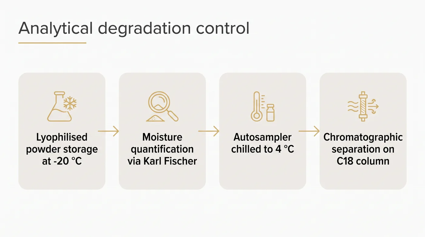
The greatest threat to a clear chromatogram is the queue. Liquid NAD+ samples left waiting in a room-temperature autosampler begin to degrade rapidly. The water hydrolyses the molecule, shrinking the primary peak and spawning new impurity signals on the readout. If a sample sits out for twelve hours, the measured purity drops artificially. Reliable testing facilities solve this by keeping their autosampler trays chilled to 4 °C, freezing the chemical clock while the instrument works through the batch.
Mass spectrometry and the sodium adduct trap
When a laboratory needs absolute confirmation of the molecule's identity, they turn to liquid chromatography coupled to tandem mass spectrometry (LC-MS/MS). The instrument vaporises the sample, applies a positive charge via electrospray ionisation, and hurls the fragments at a detector. The signature precursor ion for the NAD+ free acid appears consistently as the [M+H]+ peak at an m/z of 664. Fragmentation of this specific peak yields the distinct product ions used to build a reliable standard curve.
Mass spectrometry comes with its own specific illusions. Spectra of the free acid frequently show a secondary peak at an m/z of 686. This is a sodium adduct, a temporary pairing of the NAD+ molecule with a stray sodium ion. An inexperienced analyst looking at that 686 peak will confidently declare the powder is a sodium salt. They are usually wrong. Trace sodium leaches out of laboratory glassware and hides in mobile phase solvents, creating adducts out of thin air. An adduct on a screen proves nothing about the bulk powder in the vial.
A rigorous analytical report cuts through the noise, combining Karl Fischer moisture tracking, chilled autosamplers and clean mass spectra to deliver a verifiable result. You can view the current price, stock and detailed analytical documentation for NuuVive's NAD+ directly in the catalogue.
Questions
Why is NAD+ listed as a peptide in some catalogues?
Suppliers often group NAD+ with synthetic peptides purely for logistical convenience. It is a lyophilised powder requiring the exact same cold-chain storage and laboratory handling procedures, despite chemically being a dinucleotide coenzyme rather than an amino acid chain.
How does moisture affect the weighed mass of the powder?
The solid material is extremely hygroscopic and absorbs water from the air rapidly. If a laboratory weighs the material without subtracting the moisture content identified via Karl Fischer titration, the true concentration of the target molecule will be systematically lower than calculated.
What happens if a prepared liquid sample is left at room temperature?
Once dissolved in a liquid matrix, the molecule becomes vulnerable to hydrolytic degradation. Samples left in a warm autosampler tray will break down over several hours, which artificially lowers the measured area-percent purity on the final laboratory report.
Can a UV detector determine the salt form of the powder?
No. Standard ultraviolet detection cannot see the sodium or lithium counter-ions because they do not absorb light at the monitored wavelengths. Discovering the exact salt form requires a specific elemental analysis or reliance on the manufacturer's original specification.